NEET Test Series from KOTA - 10 Papers In MS WORD
WhatsApp Here
BIOXI20: LOCOMOTION AND MOVEMENT
340103
Which muscle go into fatigue?
1 Striped
2 Unstriped
3 Cardiac
4 Involuntnary
Explanation:
Skeletal muscle(striped muscles) get fatigue.
BIOXI20: LOCOMOTION AND MOVEMENT
340104
The H-Zone in the skeletal muscle fibre is due to
1 The central gap between actin filaments extending through myosin filaments in the A-band
2 Extension of myosin filaments in the central portion of the A-band.
3 The absence of myofibrils in the central portion of A-band
4 The central gap between myosin filaments in the A-band.
Explanation:
As per sliding filament theory, during muscle contraction, actin and myosin filaments slide over each other by formation of cross bridges and reduce length of a sarcomere. As the muscle contracts, Z-lines come closer, width of 'I' band and ' H 'band decreases. But there is no change in length of ' A ' band.
AIPMT - 2013
BIOXI20: LOCOMOTION AND MOVEMENT
340105
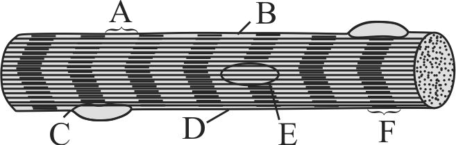
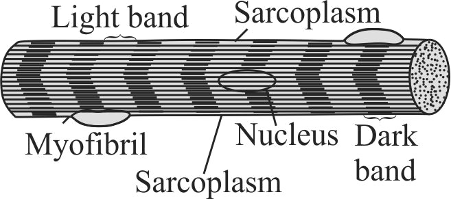
The diagram given below represents the histology of a striped muscle. Label the parts \(\mathrm{A}, \mathrm{B}, \mathrm{C}, \mathrm{D}, \mathrm{E}\) and \(\mathrm{F}\)
1 A - Sarcoplasm, B - Nucleus, C - Sarcolemma, D - Myofibril, E - Dark band , F - Light band
2 A - Sarcoplasm, B - Light band, C - Myofibril, D - Sacrolemma, E - Nucleus, F - Dark Band
3 A - Light Band, B - Sarcoplasm, C - Myofibril, D - Sacrolemma, E - Nucleus, F - Dark band
4 A - Sacrolemma, B - Nucleus, C - Dark Band, D - Light Band, E - Sacroplasm, F - Myofibril
Explanation:
KCET - 2011
BIOXI20: LOCOMOTION AND MOVEMENT
340106
Each organised skeletal muscle in our body is made of a number of
1 Muscle bundles
2 Fascicles
3 Both 1 and 2
4 Sarcomere
Explanation:
Each organised skeletal muscle in our body is made of a number of muscle bundles or fascicles held together by a common collagenous connective tissue layer called fascia.
340104
The H-Zone in the skeletal muscle fibre is due to
1 The central gap between actin filaments extending through myosin filaments in the A-band
2 Extension of myosin filaments in the central portion of the A-band.
3 The absence of myofibrils in the central portion of A-band
4 The central gap between myosin filaments in the A-band.
Explanation:
As per sliding filament theory, during muscle contraction, actin and myosin filaments slide over each other by formation of cross bridges and reduce length of a sarcomere. As the muscle contracts, Z-lines come closer, width of 'I' band and ' H 'band decreases. But there is no change in length of ' A ' band.
AIPMT - 2013
BIOXI20: LOCOMOTION AND MOVEMENT
340105
The diagram given below represents the histology of a striped muscle. Label the parts \(\mathrm{A}, \mathrm{B}, \mathrm{C}, \mathrm{D}, \mathrm{E}\) and \(\mathrm{F}\)
1 A - Sarcoplasm, B - Nucleus, C - Sarcolemma, D - Myofibril, E - Dark band , F - Light band
2 A - Sarcoplasm, B - Light band, C - Myofibril, D - Sacrolemma, E - Nucleus, F - Dark Band
3 A - Light Band, B - Sarcoplasm, C - Myofibril, D - Sacrolemma, E - Nucleus, F - Dark band
4 A - Sacrolemma, B - Nucleus, C - Dark Band, D - Light Band, E - Sacroplasm, F - Myofibril
Explanation:
KCET - 2011
BIOXI20: LOCOMOTION AND MOVEMENT
340106
Each organised skeletal muscle in our body is made of a number of
1 Muscle bundles
2 Fascicles
3 Both 1 and 2
4 Sarcomere
Explanation:
Each organised skeletal muscle in our body is made of a number of muscle bundles or fascicles held together by a common collagenous connective tissue layer called fascia.